
Home /Company News /Blog /What Is a Radiolucent ECG Electrode — and Why Does It Matter in Imaging and Cath Lab Settings? /
What Is a Radiolucent ECG Electrode — and Why Does It Matter in Imaging and Cath Lab Settings?
2026-10-30
Part 1: Why Standard Electrodes Are Radio-Opaque
A standard ECG electrode uses an ABS base with a silver/silver chloride (Ag/AgCl) conductive layer. This design has been the clinical standard for decades — it delivers excellent signal quality, stable impedance, and reliable performance across the vast majority of monitoring scenarios.
But there is one clinical scenario where the metallic component becomes a consideration: when the patient needs X-ray imaging at the same time.
Metal blocks X-rays. When a patient wearing standard ECG electrodes undergoes fluoroscopy, CT, or X-ray imaging, the metallic components can create artifacts on the image — shadows or streaks that may obscure the underlying anatomy.
This doesn't mean standard electrodes are "bad". It means that in specific imaging-intensive environments, a different design may be more appropriate.
Part 2: How Radiolucent Electrodes Work
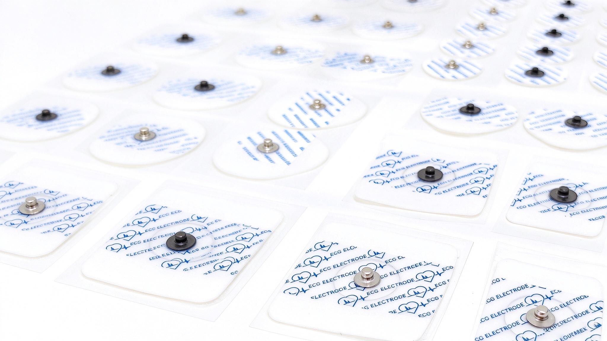
A radiolucent ECG electrode is designed to be transparent to X-rays while still providing reliable ECG signal acquisition. But how does it achieve this — and what’s different inside?
The physics of artifacts
A standard electrode contains metallic components — the sensor and backing. When X-rays pass through the body, these metal parts absorb or scatter the radiation, creating shadows and streaks on the image. In fluoroscopy and CT, these artifacts can obscure the exact anatomy the clinician is trying to see.
How radiolucent electrodes solve it
A radiolucent electrode replaces metallic components with materials that have a low atomic number — meaning X-rays pass through them almost as easily as they pass through human tissue.
A standard ECG electrode uses an ABS base with a silver/silver chloride (Ag/AgCl) conductive layer. This design has been the clinical standard for decades — it delivers excellent signal quality, stable impedance, and reliable performance across the vast majority of monitoring scenarios.
But there is one clinical scenario where the metallic component becomes a consideration: when the patient needs X-ray imaging at the same time.
Metal blocks X-rays. When a patient wearing standard ECG electrodes undergoes fluoroscopy, CT, or X-ray imaging, the metallic components can create artifacts on the image — shadows or streaks that may obscure the underlying anatomy.
This doesn't mean standard electrodes are "bad". It means that in specific imaging-intensive environments, a different design may be more appropriate.
Part 2: How Radiolucent Electrodes Work
A radiolucent ECG electrode is designed to be transparent to X-rays while still providing reliable ECG signal acquisition. But how does it achieve this — and what’s different inside?
The physics of artifacts
A standard electrode contains metallic components — the sensor and backing. When X-rays pass through the body, these metal parts absorb or scatter the radiation, creating shadows and streaks on the image. In fluoroscopy and CT, these artifacts can obscure the exact anatomy the clinician is trying to see.
How radiolucent electrodes solve it
A radiolucent electrode replaces metallic components with materials that have a low atomic number — meaning X-rays pass through them almost as easily as they pass through human tissue.
| Component | Standard Electrode | Radiolucent Electrode |
| Conductive element | ABS + silver/silver chloride (Ag/AgCl) | Carbon-based material + silver/silver chloride (Ag/AgCl) |
| Backing | Foam or non-woven | Foam or non-woven |
Key difference: The standard electrode uses an ABS base with an Ag/AgCl conductive layer. The radiolucent electrode replaces the ABS base with a carbon-based material, while keeping the Ag/AgCl conductive layer. This reduces the metallic components that create X-ray artifacts, making the electrode less visible under X-ray.
The engineering challenge is not just "removing the metal". It's maintaining electrical performance — stable impedance, reliable signal capture, secure adhesion — while making the electrode less visible to X-rays. Carbon-based conductive materials must match the signal quality of their metallic counterparts, and the adhesive must hold securely throughout long procedures.
The result
In imaging-intensive environments — catheterization labs, interventional radiology, electrophysiology labs — the ability to monitor the heart without compromising image quality is not a preference. It's a requirement.
This is where a radiolucent electrode becomes not just an option, but the right tool for the job.
Part 3: Where Radiolucent Electrodes Matter Most
The engineering challenge is not just "removing the metal". It's maintaining electrical performance — stable impedance, reliable signal capture, secure adhesion — while making the electrode less visible to X-rays. Carbon-based conductive materials must match the signal quality of their metallic counterparts, and the adhesive must hold securely throughout long procedures.
The result
In imaging-intensive environments — catheterization labs, interventional radiology, electrophysiology labs — the ability to monitor the heart without compromising image quality is not a preference. It's a requirement.
This is where a radiolucent electrode becomes not just an option, but the right tool for the job.
Part 3: Where Radiolucent Electrodes Matter Most
| Clinical Setting | Why Radiolucent Electrodes Are Used |
| Cardiac catheterization lab | Fluoroscopy is used continuously during procedures; radiolucent electrodes minimize interference with imaging |
| Interventional radiology | CT or fluoroscopy guidance requires clear images; electrode artifacts can obscure target areas |
| Electrophysiology (EP) lab | Mapping and ablation procedures rely on precise imaging |
| Hybrid OR | Combined surgical and imaging procedures; ECG monitoring must not compromise image quality |
| CT-guided procedures | ECG gating is sometimes needed during CT; standard electrodes may create streak artifacts |
Part 4: Performance Considerations
Both standard and radiolucent electrodes are engineered to meet the same electrical performance standards:
Part 5: Selection Considerations
When choosing between standard and radiolucent electrodes, consider:
Final Takeaway
Standard and radiolucent ECG electrodes serve different clinical needs. Standard electrodes remain the default choice for routine monitoring. Radiolucent electrodes are designed for imaging-intensive environments where clear visualization is essential.
At APK, what we offer goes beyond accessories — we provide reliable solutions tailored to every clinical setting.
Both standard and radiolucent electrodes are engineered to meet the same electrical performance standards:
| Parameter | Requirement |
| Impedance | Low and stable |
| Adhesion | Secure for procedure duration |
| Signal quality | Clear ECG trace, minimal noise and motion artifact |
| Biocompatibility | Meets ISO 10993 requirements |
When choosing between standard and radiolucent electrodes, consider:
- Clinical setting — is X-ray imaging performed simultaneously with ECG monitoring?
- Procedure duration — longer procedures require adhesives that maintain contact over time
- Imaging modality — fluoroscopy, CT, and X-ray have different artifact sensitivities
- Patient population — pediatric and adult patients have different size requirements
- Compatibility — electrodes must work with existing lead wires and monitor connections
Final Takeaway
Standard and radiolucent ECG electrodes serve different clinical needs. Standard electrodes remain the default choice for routine monitoring. Radiolucent electrodes are designed for imaging-intensive environments where clear visualization is essential.
At APK, what we offer goes beyond accessories — we provide reliable solutions tailored to every clinical setting.
Explore life signal, benefit universal health.
---------------------------------------------------------
APK Technology, a national high-tech enterprise with over 20 years of professional technologies and experience in providing OEM/0DM services for world-leading monitoring and ECG brands, delivering high-quality solutions for the global healthcare industry.
---------------------------------------------------------
APK Technology, a national high-tech enterprise with over 20 years of professional technologies and experience in providing OEM/0DM services for world-leading monitoring and ECG brands, delivering high-quality solutions for the global healthcare industry.
Learn more about:
Understanding "Low Perfusion": Why SpO₂ Sensor Accuracy Matters
How to Choose an ECG Electrode — A Practical Selection Guide
Related Article
ECG electrodes are the first point of contact between the patient and the monitoring system. They pick up the heart's electrical signal and pass it on to the lead wires, cables, and eventually the monitor.
But not all electrodes are the same. They vary by conductive element, backing material, shape, size, and whether they come with a pre-attached leadwire. Choosing the right one depends on the clinical setting, the patient population, and the duration of monitoring.
How to Choose an ECG Electrode — A Practical Selection Guide

In our last edition, we explored a question that most people never think to ask: when an ECG report says "Normal", how reliable is that conclusion?
The answer came down to signal quality — and the chain of components that carry the heart's electrical signal from the patient to the monitor.
How to Read an ECG Waveform — A Practical Guide for Clinicians and Engineers

You've just finished your annual health check. The doctor hands you a stack of reports. You flip to the ECG page, and there it is — a long strip of squiggly lines, a few numbers, and a single line at the bottom:"Sinus rhythm. Normal ECG."
You close the report and move on with your day.
But have you ever wondered — what does that line actually mean? And could a "normal" reading ever be wrong?
You close the report and move on with your day.
But have you ever wondered — what does that line actually mean? And could a "normal" reading ever be wrong?
Your ECG Report Says "Normal" — But Is That Conclusion Reliable?